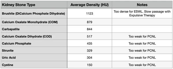
Understanding the composition and density of kidney stones plays a key role in determining the most appropriate treatment. As we discussed in our previous blog (Kidney Stone Imaging Techniques), Computed Tomography (CT) is widely used to examine stones in the urinary system. CT scans are currently the gold standard of care when it comes to kidney stone imagining because in addition to providing the size and location of the kidney stone, CT scans can also assess the density of kidney stones in Hounsfield Units (HU).
WHAT ARE HOUNSFIELD UNITS?
Sir Godfrey Newbold Hounsfield first introduced the principle of quantifying the amount of X-rays that pass through or are absorbed by tissues to create the first radio density scale. CT scan images are made from multiple x-ray images that contain pixels. Each of these pixels is assigned a gray scale value from 1 (black) to 256 (white). These values correspond to the amount of X-rays that pass through the structure and can be expressed in Hounsfield Units (HU).
The radio density of water is assigned a value of 0. Fat has a negative HU value and blood and other tissues have positive HU. For additional reference, air is assigned an HU value of -1000 and bone has an HU value of 1000. Using this method it is possible to differentiate the 256 shades of grey in a CT scan image that would be otherwise indistinguishable to the naked eye.
SIGNIFICANCE OF HOUNSFIELD UNITS
Hounsfield Units have been used in urology to predict the type and opacity of kidney stones during diagnosis. Additionally, they have been used to assess the varying treatment options available to kidney stone formers: Expulsive Therapy (Natural- like CLEANSE or Medical), Extracorpeal Shock Wave Lithotripsy (EWSL), Percutaneous Nephrolithotomy (PCNL), and Ureterorenoscopic Ureterolithotripsy (URSL).
A few notes with regards to the Hounsfield Unit density and performance of treatment options:
- Extracorpeal Shock Wave Lithotripsy (ESWL): Successful fragmentation decreased in stones with HU > 1000
- Percutaneous Nephrolithotomy (PCNL): Stones with HU < 675 reduced success of procedure by 2.65x (less dense stones fractured upon exit during surgery causing complications)
- Ureterorenoscopic Ureterolithotripsy (URSL): No impact on success of treatment
- Expulsive Therapy (NET or MET): Stones with HU > 1000 demonstrated marked slower passage due to compact structure.

As mentioned in our previous blog (Kidney Stone Imaging Techniques), we recommend that first time kidney stone formers choose to get a CT Scan. This will provide the first time kidney stone former with exceptionally valuable information about their stone. They walk away with stone size (length, width, and diameter), stone location, hydronephrosis status, and stone density information. This data will set them up for success with their current kidney stone and future kidney stones.
For future imaging needs for recurrent kidney stones, we recommend Ultrasonography. There’s no radiation exposure as with CT scans and you can still get valuable information such as stone size, stone location, and hydronephrosis status. Kidney stone types very rarely change for individuals, so the stone density information that you obtained during your initial scan for your first kidney stone is still valid. Thus, no need for consistent CT scans.
References


Why low density stone weak for pcnl, and what is the density of matrix stone
Hey Sanjay, can you rephrase your question. Not quite sure how to answer you.
Hi Sanjay, PCNL is specially designed for large, complicated stones that are not passable. Weak-density stones don’t need PCNL as they generally break apart and pass regardless of the size.
When it comes to matrix stones, do you mean mixed stones?